


Murine model for studying periodontitis
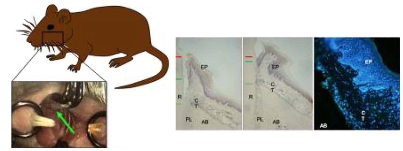
Porphyromonas gingivalis ligature-induced periodontitis
Experimental induction of periodontitis. Ligatures infected with P. gingivalis were placed along the cervical margins on the palatal sides of the first and second maxillary molars, as described by Saadi-Thiers et al. in J. Periodontol. 84, 396–406 (2013).
Infection of spheroids
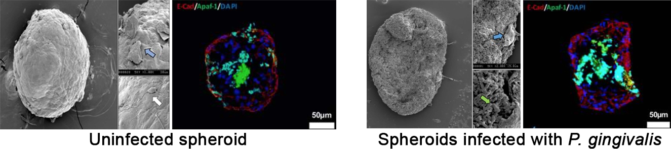
P. gingivalis bypasses the epithelial barrier to reach the fibroblasts (FB) at the core of the mucosal tissue (MT), leading to MT destruction. Scanning electron microscopy (SEM) imaging of uninfected MT and MT infected with P. gingivalis for 24 hours. Green arrows indicate P. gingivalis infection in the MT and/or cellular invasion; blue arrows indicate disruption of the epithelial barrier, multiple apoptotic cells and acute cellular damage across all layers of the MT — epithelial cells (EC) and FB; the white arrow indicates EC proliferation on the MT surface. Immunofluorescence staining of uninfected and P. gingivalis-infected MT for 24 hours (green: APAF-1, red: E-cadherin, blue: DAPI nuclear staining).
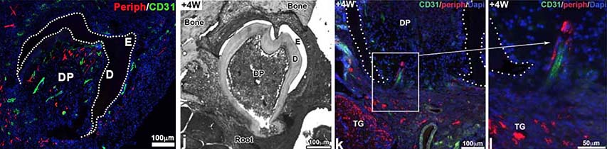
Vascularization and innervation of a biofabricated tooth
Bone/tooth interaction and eruption
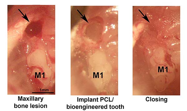
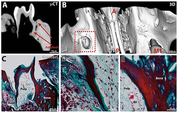
Observation of an erupted bioengineered tooth. Micro-computed tomography (µCT, A), 3D reconstruction (B) and histology (C–E, Gömöri trichrome stain) of a biofabricated tooth implanted with a PCL membrane for 10 weeks in the maxillary bone. PDL = periodontal ligament.
Scientific expertise
- Development of innovative pharmacological treatment of periodontal and oral bony defects,
- Assessment of the pathophysiological processes involved in periodontitis and development of innovative therapies for periodontal regeneration,
- Engineering of the dental organ: coronary morphogenesis, vascularization, innervation, eruption and bone/tooth interactions.